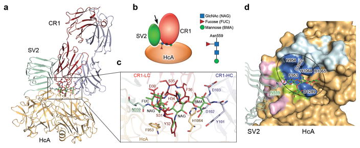
Figure 5.
The SV2 glycan-binding site on BoNT/A1 is the target for the neutralizing antibody CR1. (a,b) BoNT/A1-neutralizing therapeutic antibody CR1 (PDB code: 2NYY) 29 occupies the SV2C glycan-binding site on HCA, but does not affect HCA’s SV2C peptide-binding site. An arrow in the schematic diagram indicates the side-to-side clash between SV2C and CR1. (c) A close-up view of the interface. gSV2C-N559 glycan (green), HCA-F953 and -H1064 (gold), residues 30–36 of CR1 ligand chain (LC, red), and residues 101–103 of CR1 heavy chain (HC, gray) are shown in stick models. (d) Residues of HCA that exclusively bind SV2C peptide, N559 glycan or CR1 are colored in purple, green or cyan, respectively, while the HCA residues that are contacted by both N559 glycan and CR1 are in blue.

