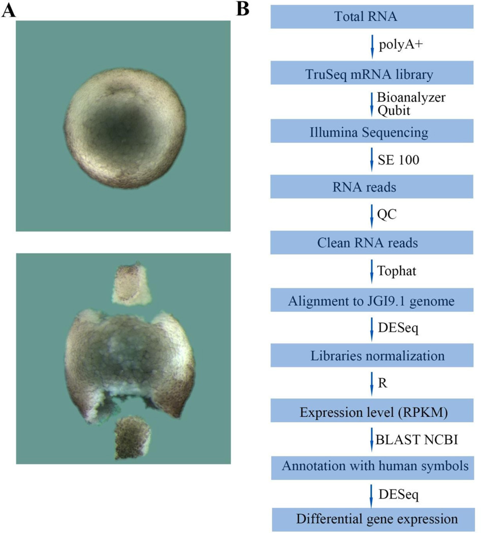
Fig. 1.
D-V dissections and experimental flowchart. (A) Dorsal and ventral lip dissection. The upper panel shows a wild-type stage 10.5 Xenopus embryo, while the lower panel shows dorsal (Dlip, top) and ventral lips (Vlip, bottom) dissected from a sibling stage 10.5 embryo. (B) Outline of the RNA-seq analysis pipeline described in materials and methods.

