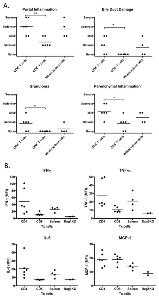
Figure 7.
Liver histopathology and level of inflammatory cytokines in recipient mice. 7A. Pathological score of portal inflammation, bile duct damage, granuloma and parenchymal inflammation and bile duct damage in group of CD4+ T cell (n=6), CD8+ T cells (n=6) and whole spleen cells (n=4) recipient mice. 7B. Serum were collected 20 weeks after cell transfer, the levels of IFN-, TNF-α, IL-6 and MCP-1 in group of CD4+ T cell (n=6), CD8+ T cells (n=6) and whole spleen cells (n=4) recipient mice were measured by a mouse inflammatory cytokine CBA kit. *, p< 0.05; **, p<0.01, determined using Kruskal-Wallis Test (Nonparametric ANOVA). All data are representative of at least two independent experiments.

