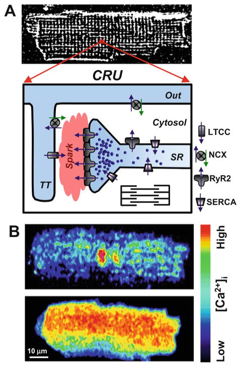
Fig. 1.
Intracellular Ca2+ regulation in adult ventricular myocytes. (a, top panel) a representative confocal image of rabbit ventricular myocytes loaded with the voltage-sensitive fluorescent dye Di-8-ANEPPS. Di-8-ANEPPS was used to label the T-tubular system. (a, bottom panel) the diagram illustrates the main components of Ca2+ release unit (CRU) in ventricular myocytes. A significant fraction of L-type Ca2+ channels (LTCC) is localized in the T-tubule (TT), whereas the majority of ryanodine receptors (RyR2) is concentrated in the junctional SR. Ca2+-ATPase (SERCA) pumps cytosolic Ca2+ back into the SR, and the Na+-Ca2+ exchanger (NCX) removes Ca2+ from the cell. The plasmalemmal Ca2+-ATPase and mitochondria play a minor role in cardiac relaxation. (b) Confocal images of diastolic Ca2+ spark (top) and an action potential-induced Ca2+ transient (bottom). Activation of a single CRU generates a Ca2+ spark, whereas simultaneous activation of thousands of these individual release units generates a global Ca2+ transient

