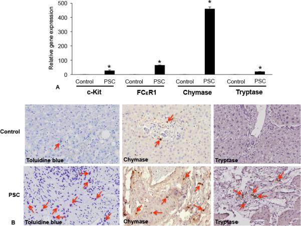
Figure 2.

Mast cell presence was assessed in human liver biopsy samples from control (no disease) and PSC patients (late and advanced PSC) by real-time PCR, toluidine blue staining and immunohistochemistry for mast cell markers (chymase and tryptase). (A) The gene expression of c-Kit, FCεR1, chymase and tryptase increased in samples from advanced and late stage PSC when compared to normal, non-diseased tissues. (B) By immunostaining (toluidine blue) and immunohistochemistry (chymase and tryptase), there is an infiltration of mast cells found surrounding damaged bile ducts in PSC patients compared to normal tissue (red arrows depict mast cells). Data are expressed as mean ± SEM of at least 6 experiments for real-time PCR. *p<0.05 versus control. Images are 20× magnification
