Figure 5.

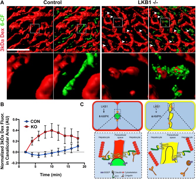
IVM of TJs permeability and the paracellular pathway. (A) Mice were anesthetized and positioned on the microscope stage for IVM of the liver as described in the Materials and Methods section. Blood flow and sinusoid space were visualized with a Qtracker 655 probe (not shown) that was injected systemically prior to imaging. 3 kDa dextran and 6‐CFDA were injected after acquisition of the first z‐stack. Volume rendering of the different probes was generated; a single frame of 3 kDa dextran (red) or an overlay of 3 kDa and 6‐CF (green) are shown. The enlarged inset in the lower panel corresponds to the boxed area in the upper panel. Note that in LKB1−/−, but not control, 3 kDa dextran is present in the canaliculi area also labeled by 6‐CF (white arrowheads). Scale bar: 50 μm. (B) Measurement of 3 kDa dextran in the bile canaliculi area was plotted as the mean ± SD of 20 regions for representative control mice (blue) or LKB1−/− mice (red). Similar results were obtained in three independent experiments. (C) Proposed model summarizing the experimental observations. LKB1‐deficient mice had reduced levels of phosphorylated AMPK levels, which may be the cause of the observed abnormal canaliculi morphology and distribution of TJ proteins. Notably, the apical pole extended into the lateral domain of hepatocytes and the junctional regulatory protein, cingulin (orange), localized primarily in the cytosol. Mislocalization of additional TJ proteins, and membrane trafficking regulators may explain the delayed biliary excretion. Mixing of biliary (green in control mice) and blood (red in control mice) content (represented as yellow) was observed in LKB1‐deficient mice.
