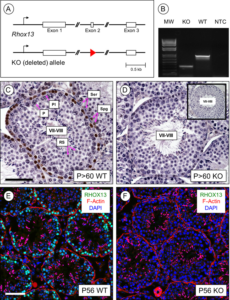
Figure 1. Generation of Rhox13 KO mice.
(A) The Rhox13 gene is comprised of three exons (shown in boxes), as is typical of most homeobox genes. We created a KO allele by deleting exon 2, and a single loxP site remains (red triangle). (B) PCR amplification using primers on either side of exon 2 distinguishes KO from WT alleles. MW = molecular weight ladder, and NTC = no template control. (C–D) IHC was done to verify loss of RHOX13 protein (brown staining) in P>60 adult testes. Shown are stage VII-VIII seminiferous tubule sections from WT (C) and KO (D) mice. Sections were counterstained with hematoxylin, and inset (in D) is a no primary antibody control. Spg = spermatogonia, Pl = preleptotene spermatocytes, P = pachytene spermatocytes, RS = round spermatids, Ser = Sertoli cells. (E–F) IIF was done to localize RHOX13 (in green) in WT (E) and KO (F) P56 testes. DNA is stained with DAPI (blue), and F-actin is stained with fluorescently-conjugated phalloidin (red). Scale bar = 50 µm.

