
Figure 5.
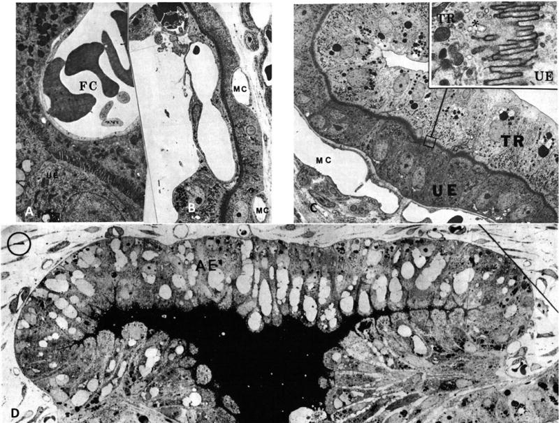
The porcine placenta: A, Fetal capillaries (FC) at d 58 of pregnancy protrude deeply into the trophoblast, with the diffusion distance between fetal bloodstream and maternal uterine epithelium (UE) reduced to a few μm; B, At d 110 of pregnancy maternal capillaries (MC) project between uterine epithelial cells bringing the maternal and fetal capillaries within 3–5 μm; C, At d 30 of pregnancy, the microvilli on the trophoblast surface (TR) interdigitate with ones on the uterine epithelium (UE) to provide an intimate contact layer. Maternal capillaries (MC) are placed just below the basal lamina of the UE; D, A general view of a dome-shaped areola (AE) situated above the mouth of a uterine gland (UG) at d 30 of pregnancy. Figures 5A-C are from Friess et al. (1980) (Friess, et al. 1980); Figure 5D is from Friess et al. (1981)(Friess, et al. 1981) with permission.