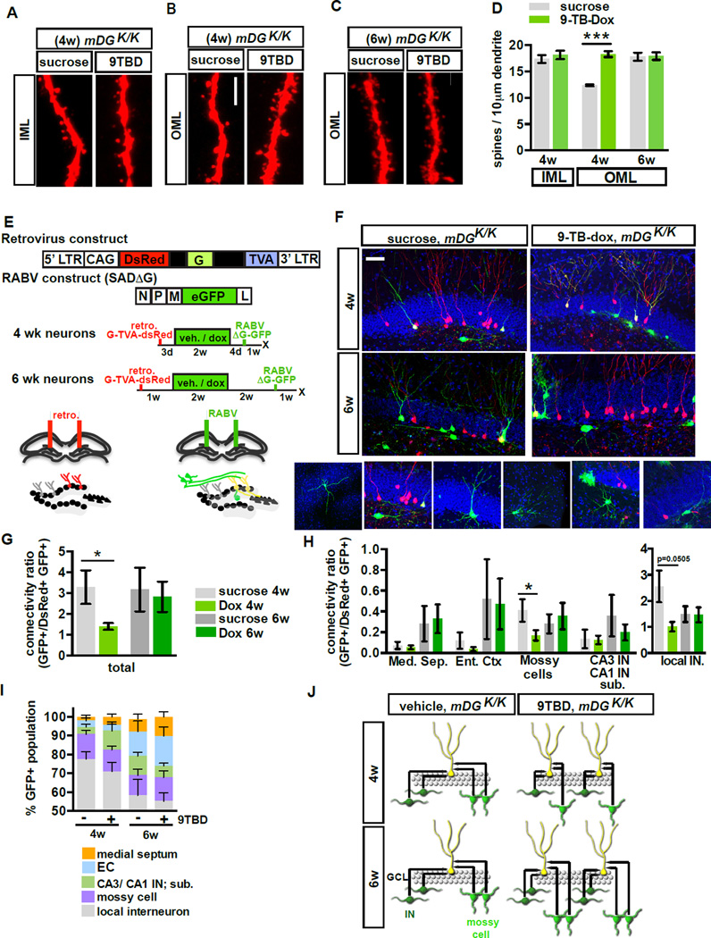
Figure 5. Genetic enhancement of adult hippocampal neurogenesis transiently reorganizes local afferent connectivity of maturing adult-born DGCs.
A - D) 4 but not 6-week-old adult-born DGCs of 9TBD-treated mDGK/K mice have greater spine density than vehicle treated mDGK/K mice. (A, C) Maximum intensity projections of confocal z-stack scans of OML and (B) IML dendritic segments 4 weeks (A,B) or 6 weeks (C) after injection of DsRed-expressing retrovirus. 9TBD was administered as shown in Fig. 5E. (D) Quantification of A-C (4wk n=5,4, 6w n=3,3).
E - J) Decreased local monosynaptic inputs onto 4-week-old, but not 6-week-old, adult-born DGCs in 9TBD treated mDGK/K mice. (E) Schematic showing rabies virus constructs and injection paradigm for F-J. (F) Representative images of dorsal DG sections showing retrovirally labeled cells (red), starter cells (yellow), and presynaptic partners (green). Below, images of specific presynaptic cell types. (G) Connectivity ratio of 4-week, but not 6-week-old, adult-born DGCs is decreased in 9TBD-treated mDGK/Kmice relative to controls (n=4 for all groups) with (H) specific reductions in connectivity to mossy cells and interneurons (n=4 DG INs p=0.0505). (I) 4 and 6-week-old adult-born DGCs of vehicle and 9TBD treated mDGK/K show similar distributions of pre-synaptic cells. (J) Population-level model conveying how enhancing adult hippocampal neurogenesis reorganizes inputs from mossy cells and DG INs. Scale bar: 5µm (A,C), 20 µm (F).

