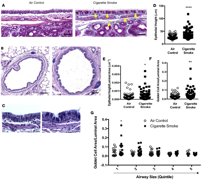
Figure 2. Histopathologic evidence of chronic mucus hypersecretion and goblet cell hyperplasia in a ferret model of COPD.
(A) Mucus-expressing goblet cells and submucosal glands (yellow arrows) were stained with PAS (magenta) in tracheal sections of ferrets exposed to room air or cigarette smoke for 6 months. Scale bar: 34 μm. (B) Similarly, lung sections from the same ferrets were stained with AB-PAS to highlight mucus-expressing cells (deep blue). Scale bar: 110 μm. (C) The same staining as in B demonstrating goblet cell hyperplasia and increased epithelial cell height. Scale bar: 17 μm. (D and E) Quantitative analyses demonstrating epithelial hyperplasia, as measured by increased epithelial cell height (D) controlled for luminal area (E). (F) Goblet cell area controlled for luminal area in cigarette smoke–exposed ferrets. (G) Goblet cell area controlled for luminal area expressed as a function of the airway luminal (inner) diameter quintile (diameter; quintile 1: 116–293 μm, quintile 2: 294–415 μm, quintile 3: 424–535 μm, quintile 4: 537–793 μm, and quintile 5: 810–2,651 μm). n = 8/group. *P < 0.05, **P < 0.01, ****P < 0.0001.

