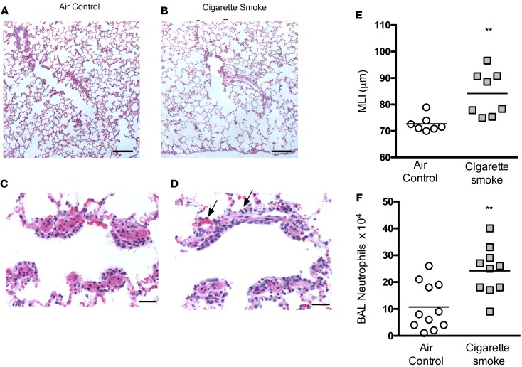
Figure 5. Emphysema and neutrophilic inflammation in smoke-exposed ferrets.
(A and B) Histopathologic evidence of emphysematous airspace enlargement shown in representative lung sections of ferrets exposed to cigarette smoke and their corresponding air controls. Scale bar: 220 μm. (C and D) H&E-stained section of respiratory bronchioles, indicating an influx of neutrophils (black arrows) in ferrets exposed to cigarette smoke for 6 months compared with air controls. Scale bar: 34 μm. (E) Summary of mean linear intercept (MLI), a marker of alveolar enlargement, in ferrets exposed to cigarette smoke or air controls for 6 months. (F) Summary of mean neutrophil counts observed in bronchoalveolar lavage fluids in cigarette smoke–exposed ferrets and their air control counterparts. n = 8–12/group. **P < 0.01.

