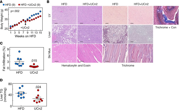
Figure 7. Weight gain and fatty infiltration of liver.
(A) Weight gain on HFD. Weights increased similarly in both groups for 8 weeks but were attenuated after UCn2 gene transfer (P = 0.002; repeated measures ANOVA). (B) Histology of liver and left ventricle. Mice fed HFDs for 5 weeks received i.v. UCn2 gene transfer (HFD + UCn2; 5 × 10¹¹ gc) or saline (HFD), and HFD was maintained for 5 additional weeks. AAV8.UCn2 delivery and sustained urocortin 2 expression had no adverse effects on liver, LV, or skeletal muscle histology. Evident in the liver micrographs is fatty infiltration in HFD-fed but untreated mice. H&E and Masson’s trichrome staining were performed on sections of liver, transmural sections of left ventricle (LV), and skeletal muscle (Skl Mus). Trichrome + Con, positive control for Masson’s trichrome stain (scale bars: 100 μm). (C) Quantitative histological assessment of liver indicates that UCn2 gene transfer was associated with a 74% reduction in fatty infiltration of liver (P = 0.015; Student’s t test, unpaired, 2-tailed). Reduced fatty infiltration of the liver appeared to be independent of weight gain; it also was seen after UCn2 gene transfer in db/db mice despite no group difference in weight. (D) Liver triglyceride content. Livers from HFD-fed mice that received UCn2 gene transfer showed reduced triglyceride content (P = 0.024; Student’s t test, unpaired, 2-tailed).

