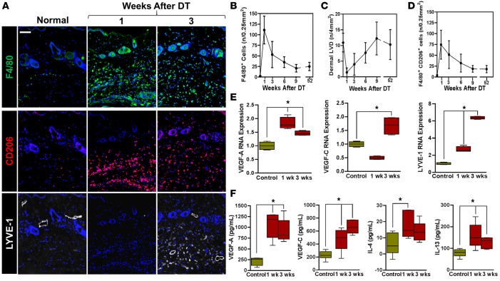
Figure 4. Lymphatic regeneration after DT administration is associated with macrophage infiltration and M2 differentiation.
(A) Representative triple immunofluorescence localization of F4/80 (top), CD206 (middle), and LYVE-1 (bottom) in the hind limbs of control (normal) and DT-treated mice 1 and 3 weeks after DT administration (scale bar: 100 μm). (B) Quantification of macrophage (F4/80+) infiltration at various time points following lymphatic ablation with DT. (C) Quantification of lymphatic vessel density over time in control and DT-treated mice. (D) Quantification of F4/80+ and CD206+ dual positive cells in hind limb tissues over time. (E) Real-time quantitative PCR for VEGF-A, VEGF-C, and LYVE-1 in cell-sorted CD11b+ cells harvested from hind limb tissues before (control) and 1 or 3 weeks after DT administration (*P < 0.01). (F) ELISA for VEGF-A, VEGF-C, IL-4, and IL-13 in hind limb tissues harvested from control and DT-treated animals 1 and 3 weeks following DT administration (*P < 0.05). 2-tailed Student’s t test.

