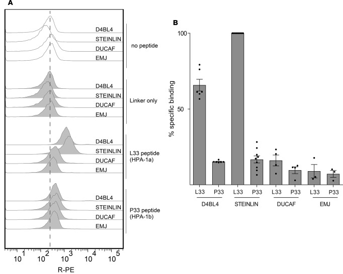
Figure 1. Peptide binding to cell lines.
Binding of control 12-mer peptides (L33 and P33) extended with a biotinylated linker peptide were incubated with DRB3*01:01-positive cell lines (STEINLIN and D4BL4) or control cell lines (DUCAF, EMJ) at 5 μM peptide in the presence of 2.5 μM AdEtOH. The efficiency of binding to APCs was assessed by flow cytometry with streptavidin-conjugated R-PE. (A) Representative raw data of one of 3 independent peptide binding assays (1 of 2 replicates shown). (B) Comparison of percent specific binding. Data points from independent experiments are presented as dots, with bars representing mean ± SEM of at least 3 experiments. Raw data values were median R-PE fluorescence intensity on B-LCLs (gated by light scatter cytogram). Background intensity (cells only, no peptide) was subtracted before calculating the specific binding within each experiment (L33 peptide on STEINLIN defined as 100%). AdEtOH, adamantane ethanol; APC, antigen presenting cell; R-PE, R-phycoerythrin; B-LCL, B-lymphoblastoid cell lines.

