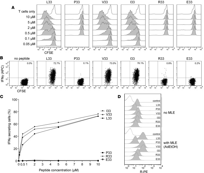
Figure 2. HPA-1a–specific T cells do not exclusively recognize the Leu33 residue in the HPA-1a peptide.
(A) CFSE-labeled HPA-1a–specific T cell clones were stimulated with DRA/DRB3*01:01-positive APCs (D4BL4) pulsed with a panel of designed peptides in different concentrations, without MLE. T cell proliferation was determined after 7 days, illustrated by 1 of 4 experiments (D7T4). (B) T cell activation was also measured by intracellular cytokine staining (IFN-γ) by flow cytometry. T cells were gated by light scatter and CFSE staining. One representative of 3 experiments with 10 μM peptide pulsing of D4BL4 cells, and (C) the effect of peptide pulsing concentrations on T cell activation. (D) Binding of peptides to D4BL4; same cell line used for T cell activation. Cells were pulsed with biotinylated peptides (10 μM) with or without AdEtOH, stained with Streptavidin–R-PE, and analyzed in flow cytometry. The discrimination of peptide-binding efficiency is enhanced when using AdEtOH as an MLE. Substitutions of Leu33 to Arg/Glu reduced binding, while efficient binding was seen with substitutions with small, aliphatic residues Leu33 to Val/Ile. Comparative binding experiments were conducted twice with 2 replicates; representative histograms are shown. Twelve-mer peptides were used in all experiments. HPA, human platelet antigen; MLE, MHC-loading enhancer; AdEtOH, adamantane ethanol.

