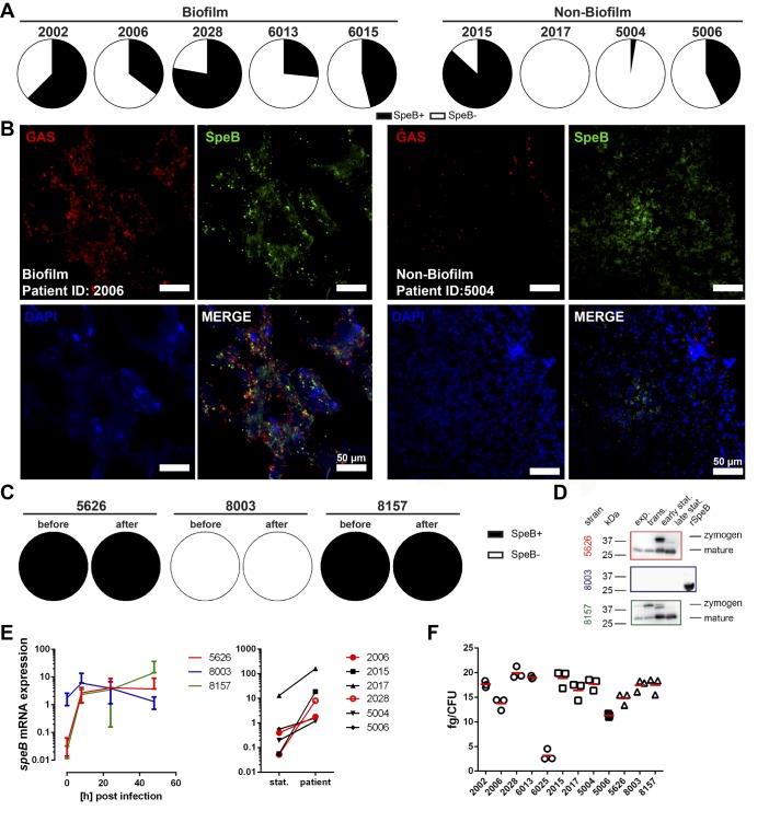
Figure 5. SpeB, capsule, and CovR/S are not associated with biofilm formation in the tissue setting.
(A) Distribution of SpeB-positive and SpeB-negative (SpeB+/SpeB–) clones recovered from biofilm- and non–biofilm-associated patient biopsies. (B) Representative immunofluorescence micrographs of the distribution of SpeB in biofilm and nonbiofilm patient biopsies (original magnification, ×40). (C) SpeB positivity among the necrotizing soft tissue infection strains used for the skin model infections (before infection and 48 hours after). (D) Western Blot analyses of secreted SpeB by indicated strains at different growth stages. Representative images of 3 experiments are shown (n = 3). (E) Relative mRNA expression of speB before and during the infection in tissue models (left panel) or in patient biopsies (right panel; stat., static culture). The data on skin tissue model (left panel) represent the mean values ± SD (n = 3). (F) Amounts of capsular hyaluronic acid in exponential growth phase bacteria. Strains from patients with biofilm-positive or -negative biopsies are shown in circles and squares, respectively. The horizontal line denotes mean values (n = 3).

