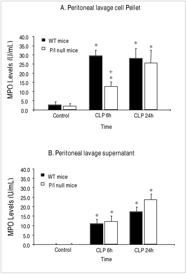
Figure 4.
Neutrophil infiltration as determined by MPO contents of peritoneal cell pellet and peritoneal supernatant. Wild-type (WT) and P/I null were subjected to CLP, euthanized at specific time points, the peritoneal lavages harvested, centrifuged, and the supernatant and the cell pellet were collected separately and assayed for MPO contents. (A) Peritoneal lavage cell pellet. (B) Peritoneal lavage supernatant. Note that at 6 h after CLP, although a significant neutrophil infiltration into the peritoneal cavity of the P/I null group is present, it is significantly impaired compared to the corresponding WT group. Values are expressed as the mean ± SEM. * p < 0.05 compared to respective control group. + p ≤ 0.05 P/I null mice compared to respective wild-type at the same time period of CLP. Data from 6 independent experiments with total sample size of 8 mice per each treatment.

