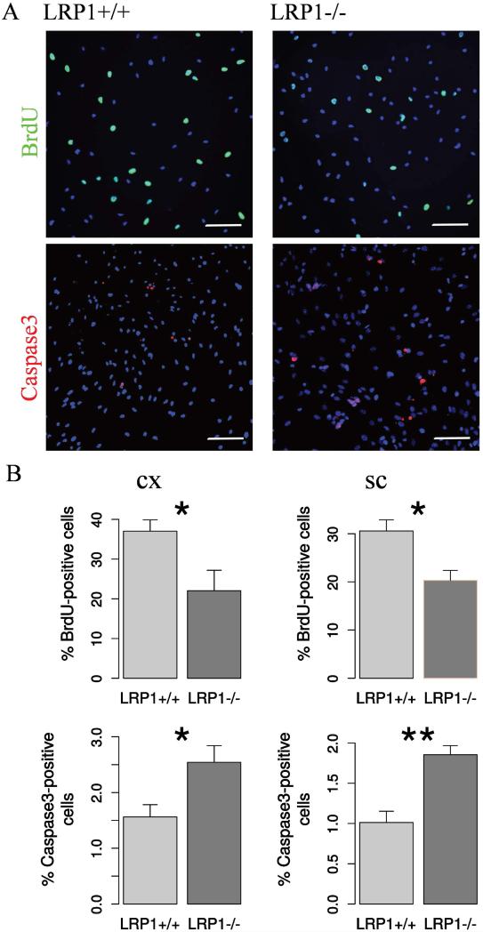
Fig. 2. LRP1 deletion interferes with NSPC proliferation and survival.
A. Representative photomicrographs of LRP1+/+ and LRP1−/− NSPCs immunostained against BrdU (N = 7, cx; N = 10, sc) and apoptosis marker active Caspase3 (N = 9, cx; N = 11, sc). Scale bar 50 μm. B. Quantification results indicate 1.5 fold reduction in proliferation and 2 fold increase in the apoptosis rate of LRP1 NSPCs derived from cortex and spinal cord. Data are expressed as mean ± SE (* indicates P < 0.05, ** — P < 0.01). Cx = cortex, sc = spinal cord.

