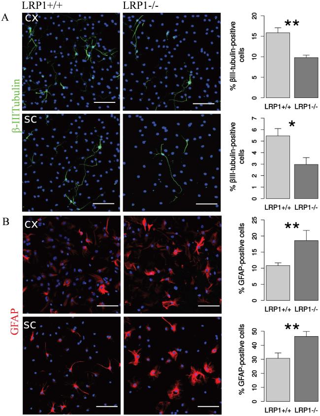
Fig. 4. LRP1 knockout NSPCs generated less neurons, but more astrocytes.
Representative photomicrographs of LRP1+/+ and LRP1−/−differentiated NSPCs derived from cortex and spinal cord immunostained against: A. βIII-tubulin — a marker of young neurons (N = 10, cx, N = 10, sc). LRP1−/− NSPCs derived from both tissues generate nearly twice less neurons. B. GFAP — an astrocytic marker (N = 7, cx; N = 14, sc). LRP1 NSPCs derived from both tissues generate around 1.5 times more astrocytes. Scale bar 50 μm. Data are expressed as mean ± SE (* indicates P < 0.05, ** — P < 0.01). Cx = cortex, sc = spinal cord.

