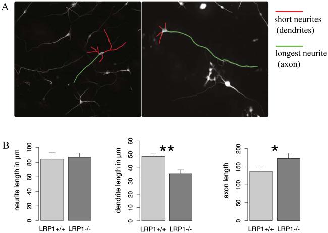
Fig. 6. LRP1 deletion modifies neuronal polarity.
A. Representative photomicrographs of LRP1+/+ and LRP1−/− differentiated NSPCs derived from cortex immunostained against βIII-tubulin. The longer neurites (presumably axons) and the shorter processes (presumably dendrites) are indicated with green and red false color traces, respectively. B. The comparison of total neurite, dendrite and axon length between LRP1+/+ and LRP1−/− neurons. LRP1 knockout neurons exhibit significantly shorter dendrites and longer axons compared to wild type neurons, indicating alterations in neuronal polarity upon LRP1 deletion.

