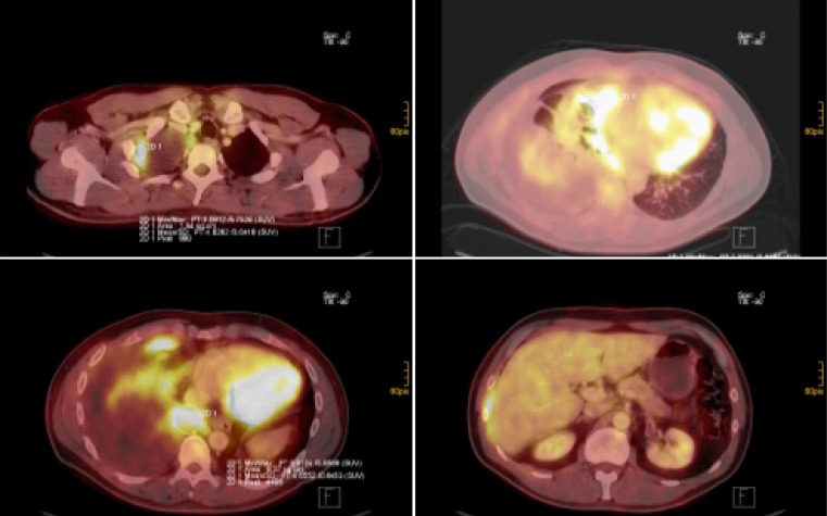
Fig. 6.
FDG PET-CT images: malignant pleural mesothelioma of the right pleural cavity (various slides of PET/CT fusion imaging). Varoius slides of CT/PET fusion imaging showing pleural tumor apical right (top left), involving the visceral and parietal pleura in the pleura costodiaphragmatic area (bottom left and right) and the pericardium (top right)

