
Fig. 5.
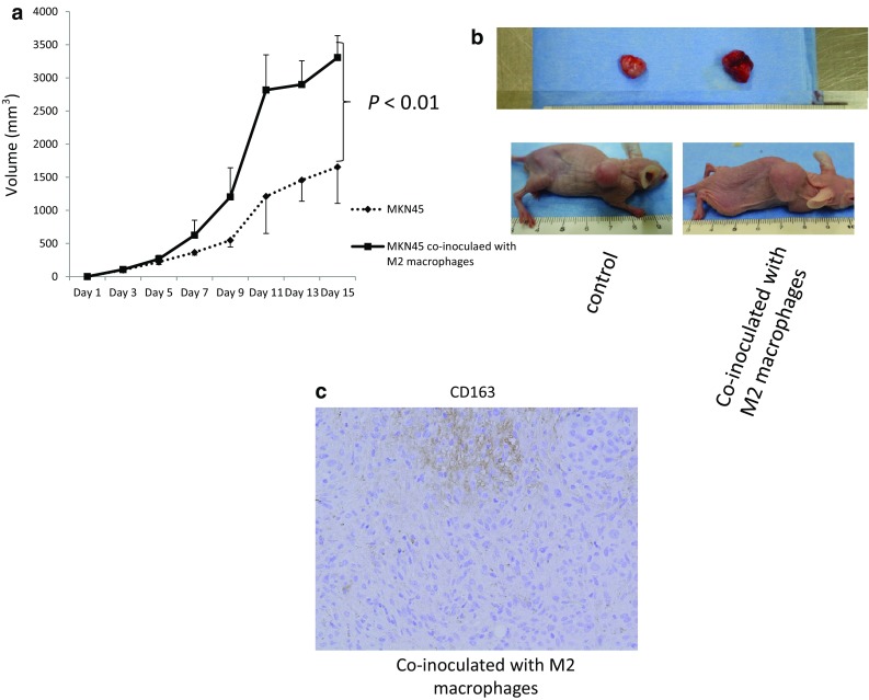
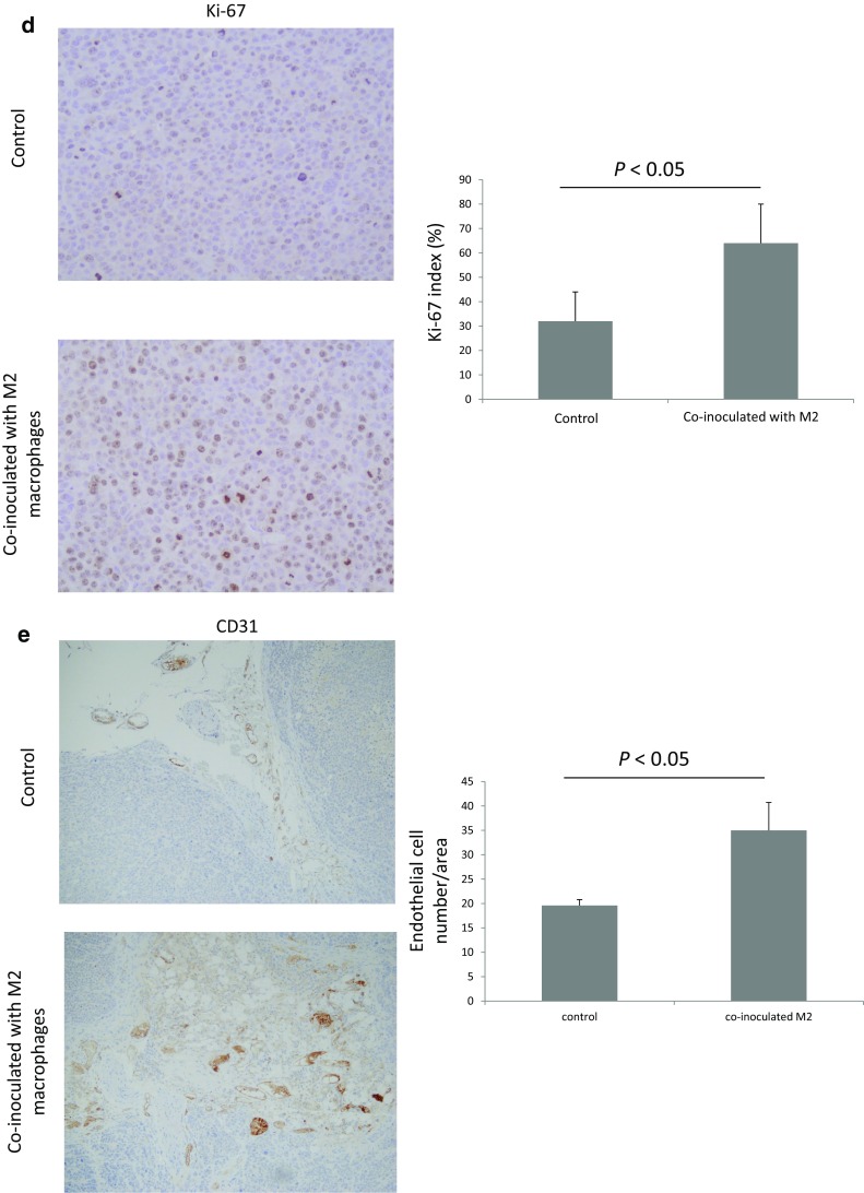
a In vivo effect of M2 macrophages on tumor growth in a xenograft model. a The mean volume of the tumors co-inoculated with MKN45 and M2 macrophages was evaluated until 15 days after inoculation. Values represent the mean ± standard error (n = 6). b Representative images showing the macroscopic appearance of the tumors at day 15. c M2 macrophages derived from peripheral blood mononuclear cells (anti-human CD163 antibody) were found in xenograft tissue. d Immunohistochemical staining with Ki-67 in the co-inoculated group and the control group inoculated with MKN45 cells alone. e Immunohistochemical staining with anti-mouse CD31 antibody. Microvessel density was evaluated as CD31+ endothelial cell number per visual field (×100)