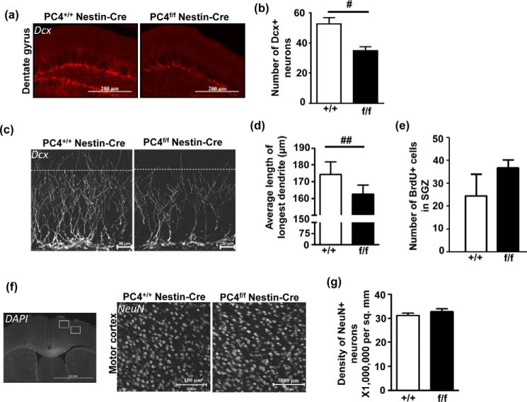
FIGURE 4.
Absence of PC4 results in altered neurogenesis. a, a typical example of the immunofluorescence analysis of Dcx (doublecortin) in cryosections. Scale bars, 200 μm. b, the number of Dcx-positive neurons in the dentate gyrus was counted in several sections per animal (n = 3; p < 0.05), and a significant decrease was observed in the number of Dcx-positive neurons in PC4f/f Nestin-Cre mice. c, typical magnified images of Dcx-stained sections showing the reduced number of neurons. The dotted line indicates a length of 100 μm from the SGZ, and most dendrites were restricted to this length in the PC4f/f Nestin-Cre mice, whereas visibly more dendrites were longer than 100 μm in the PC4+/+ Nestin-Cre mice. Scale bars, 20 μm. d, measuring the length of the longest Dcx-positive dendrite in the hippocampus showed that this length was significantly lesser in the PC4f/f Nestin-Cre mice compared with the PC4+/+ Nestin-Cre mice (PC4+/+ Nestin-Cre: 174.4 μm versus PC4f/f Nestin-Cre: 162.4 μm; n = 3; p < 0.01). e, effect of PC4 knock-out on proliferation in the SGZ after BrdU injection (100 mg/kg). Control and PC4 knock-out mice were injected with BrdU 1 h before they were sacrificed. The numbers of BrdU-positive cells in the SGZ were not statistically different between the two genotypes (n = 2–3; 4 sections/animal). f, immunofluorescence analysis of NeuN (right panel), counterstained with DAPI (left panel) in cryosections of PC4+/+ Nestin-Cre and PC4f/f Nestin-Cre mice showing density of neurons in the cortical regions (white boxes depict M1 and M2 motor cortical regions). Representative images showing NeuN-positive cells in the motor M1 cortical region are shown for both genotypes (right panel). Scale bars, 100 μm. g, the number of NeuN-positive cells in the motor cortical region was counted, and the density of neurons was seen to be similar in the PC4+/+ Nestin-Cre and PC4f/f Nestin-Cre mice (n = 3; 4–5 sections/animal).

