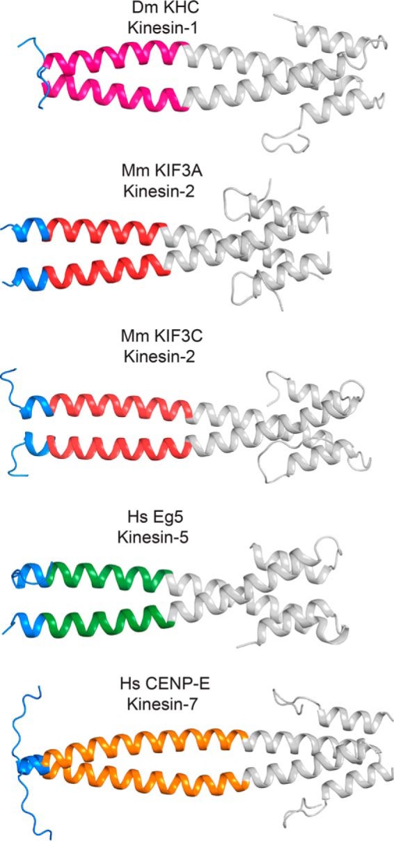
FIGURE 2.

Crystal structures of kinesin-1, -2, -5, and -7 reveal unique class-specific neck-linker α7 neck-coil domains. The neck-linker motif predicted to occur based on the earlier studies of kinesin-1 is colored blue. Helix α7, as observed in kinesin-1, is colored according to the kinesin family to which it belongs. EB1, a coiled-coil fusion protein, is colored gray. There are varied amounts of ordered neck-linker motifs. These structures show that the true start of helix α7 is variable across the kinesin superfamily. Table 4 provides the protein sequence of each fusion protein and their coiled-coil registry. Figs. 2–6 were prepared in part with PyMOL.
