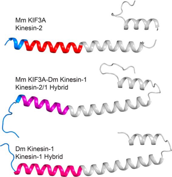
FIGURE 6.

Comparison of KIF3A, the KIF3A-kinesin-1 hybrid protein, and kinesin-1 structures. Predicted neck-linkers are shown in blue and the EB1 domain in gray. KIF3A is at the top with its helix α7 colored red, KIF3A-kinesin-1 hybrid protein in the middle with its helix α7 in purple, and kinesin-1 at the bottom with its helix α7 in hot pink. Note the variability in neck-linker length based on the start of helix α7.
