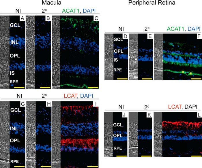
FIGURE 7.
Immunolocalizations of ACAT1 and LCAT in human retina. The left and right sections in each panel are phase contrast images and histochemistry images, respectively. A, D, G, and J, control stains with nonimmunized (NI) serum; and B, E, H, and K, secondary (2o) antibody. Nuclei were stained with DAPI (in blue). C and F, stains for ACAT1 (in green); and I and L, LCAT (in red). Immunoreactivity was detected by Alexa Fluor 647-conjugated secondary antibody. F, yellow asterisks indicate drusen. All images are representative (n = 5 donors). Sections of the macular and peripheral retina are from the same donor. Scale bars: 50 μm.

