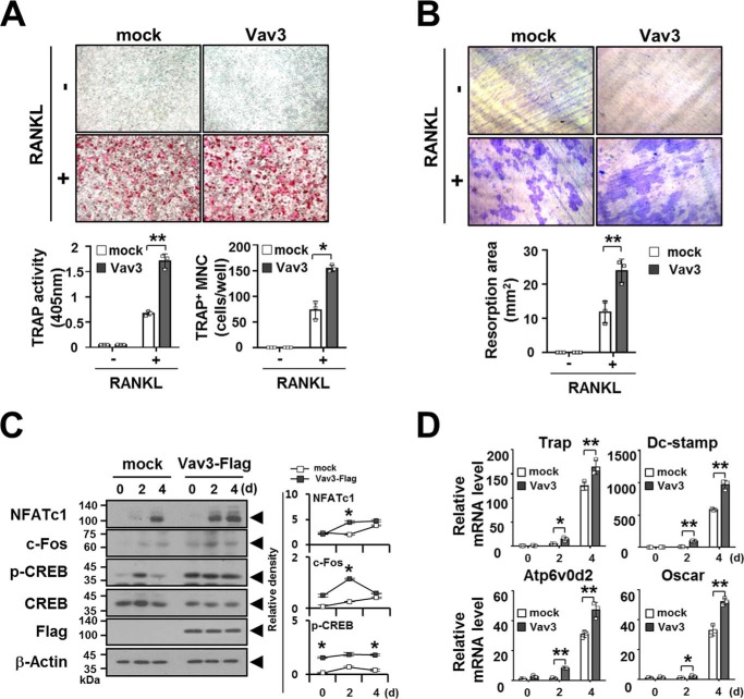
FIGURE 5.
Vav3 expression enhances osteoclastogenesis. A, the effect of Vav3 expression on OC differentiation. BMMs cultured with M-CSF (150 ng/ml) for 2 days were transduced with retroviral supernatants harboring Vav3 expression and puromycin-resistant cassettes and were further cultured with puromycin and M-CSF (150 ng/ml) for 2 days. Puromycin-selected Vav3-expressing BMMs were differentiated into OCs with RANKL (100 ng/ml) and M-CSF (50 ng/ml) for 4 days. The OCs were photographed (top panel, original magnification, ×50) after TRAP staining. The OCs were analyzed using the TRAP solution assay (bottom left panel). The number of TRAP+ MNCs (at least three nuclei) was counted (bottom right panel). mock, empty vector control. *, p < 0.05; **, p < 0.01. B, the effect of Vav3 expression on resorption pit formation. Resorption pits were visualized (top panel, original magnification, ×50). The summarized data from the resorption pit assays are shown in the bottom panel. C, the effect of Vav3 expression on the activation of osteoclastogenic factors. OCs were differentiated from BMMs with RANKL (100 ng/ml) and M-CSF (50 ng/ml) for the indicated times and subsequently analyzed via immunoblotting (IB) with antibodies that recognized phosphorylated or total NFATc1 (7A6), c-Fos (D-1), and CREB (Ser133). The cell lysates were normalized for total protein content. The level of Vav3 expression was detected via anti-FLAG IB. The relative protein level was calculated after normalization to β-actin protein input (right). The relative level of phosphorylated CREB was calculated after normalization to total CREB protein input (right). β-Actin was used as the loading control. D, the effect of Vav3 on the expression of osteoclastogenic markers. Total RNA was prepared from OCs and subjected to real time PCR analysis. The data were normalized to β-actin. All experiments were performed at least three times with similar results.

