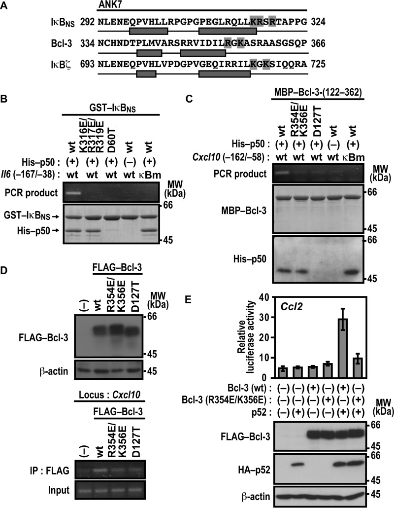
FIGURE 7.
Basic residues in ANK7 of IκBNS and Bcl-3 participate in association with their target gene promoter. A, amino acid sequence alignment of ANK7 in the mouse nuclear IκB proteins IκBζ, Bcl-3, and IκBNS. The two antiparallel α helices in ANK7 of Bcl-3 were determined in crystal structures of Bcl-3 (36), whereas those of IκBζ and IκBNS were predicted from their primary sequences by using the program PSIPRED (45, 46). Residues boxed in gray are the basic residues that follow the second α helix of ANK7. B, complex formation of IκBNS with p50 and the Il6 promoter. GST-fused WT IκBNS or a mutant IκBNS with the K316E/R317E/R319E or D60T substitution was incubated with or without His-p50 in the presence of WT Il6 (−167/−38) or a mutated κB site (κBm)-carrying Il6 (−167/−38). After the protein-DNA complex was pulled down with glutathione-Sepharose-4B beads, the co-precipitated DNA was amplified by PCR, and the product was analyzed by agarose gel electrophoresis. The precipitated proteins were subjected to SDS-PAGE, followed by staining with CBB. MW, molecular weight. C, complex formation of Bcl-3 with p50 and the Cxcl10 promoter. MBP-fused WT Bcl-3-(122–362) or a mutant protein with the R354E/K356E or D127T substitution was incubated with or without His-p50 in the presence of Cxcl10 (−162/−58) or a mutated κB site (κBm)-carrying Cxcl10 (−162/−58). After the protein-DNA complex was pulled down with amylose resins, the co-precipitated DNA was analyzed as in B. The precipitated proteins were subjected to SDS-PAGE, followed by staining with CBB or immunoblot with the anti-His antibody. D, formaldehyde-fixed chromatin was prepared from RAW264.7 cells stably expressing FLAG-Bcl-3 (WT), FLAG-Bcl-3 (R354E/K356E), or FLAG-Bcl-3 (D127T) (top panel) and subjected to ChIP assay using anti-FLAG (M2) mouse monoclonal antibody (bottom panel). Precipitated DNA was analyzed by PCR using primers corresponding to the Cxcl10 locus. The results are representative of experiments from at least three independent experiments. IP, immunoprecipitation. E, the role of Arg-354 and Lys-356 of Bcl-3 ANK7 in Ccl2 activation. RAW264.7 cells were transfected with the following plasmids: the luciferase reporter plasmid pGL3-Basic containing the upstream region of Ccl2 (−2777/+76), the internal control plasmid pRL-TK, and pcDNA3 for expression of FLAG-Bcl-3 (WT) or FLAG-Bcl-3 (R354E/K356E) and HA-p52. Luciferase activities were determined as described under “Experimental Procedures.” Each graph represents the mean ± S.D. obtained from three independent transfections. Cell lysates were analyzed by immunoblot with anti-FLAG, anti-HA, or anti-β-actin antibody. Positions for marker proteins are indicated in kilodaltons.

