FIGURE 5.
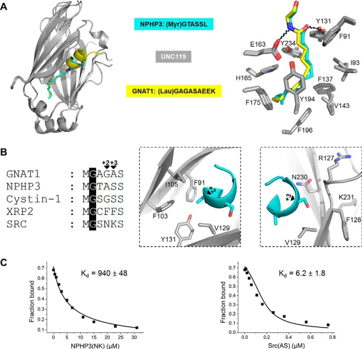
Structural analysis of a complex of Unc119a with myr-NPHP3. A, superimposition of the crystal structures of Unc119a·myr-NPHP3 and Unc119a·lau-GNAT1 (PDB code 3RBQ) with the lauroyl group in yellow and myristoyl in blue (left). The right panel shows the Unc119a residues interacting with myristoylated NPHP3 and lauroylated GNAT1. B, sequence alignment of the N-terminal part of myristoylated proteins involved in this study (left). Residues of Unc119a around the +2 (middle) and +3 (right) positions of the myr-NPHP3 are shown. C, titration of a complex between 100 nm fluorescein-labeled RP2 peptide and 200 nm Unc119a with increasing concentration of NPHP3(NK) (left) and Src(AS) (right) mutant peptides leads to a decrease in fluorescence polarization. Titration data were fitted with a competition model.

