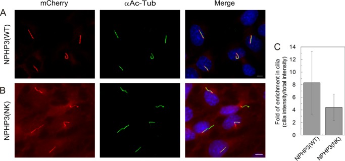
FIGURE 6.
Localization of NPHP3(WT)-mCherry and NPHP3(NK)-mCherry in IMCD3 cells. A, mCherry fluorescence (LAP-tagged) (red) shows stably expressed NPHP3(WT)-mCherry almost exclusively localizing to primary cilia, which are immunostained with an antibody against acetylated tubulin (green). B, NPHP3(NK)-mCherry localizes both to primary cilia and to the rest of the cell. White bar, 5 μm. C, ciliary enrichment of NPHP3(WT)-mCherry was compared with that of the NPHP3(NK) mutant. The bar graph shows the ratios of the mCherry fluorescence intensity in cilia relative to the total mCherry intensity outside the cilium. Ratios indicate the enrichment of mCherry-tagged NPHP3 inside the cilia. Data analysis of 40 ciliated cells each stably expressing wild type or mutant was accomplished using CellProfiler. Error bars, S.D., n = 40 (p < 0.05; Student's t test).

