FIGURE 5.

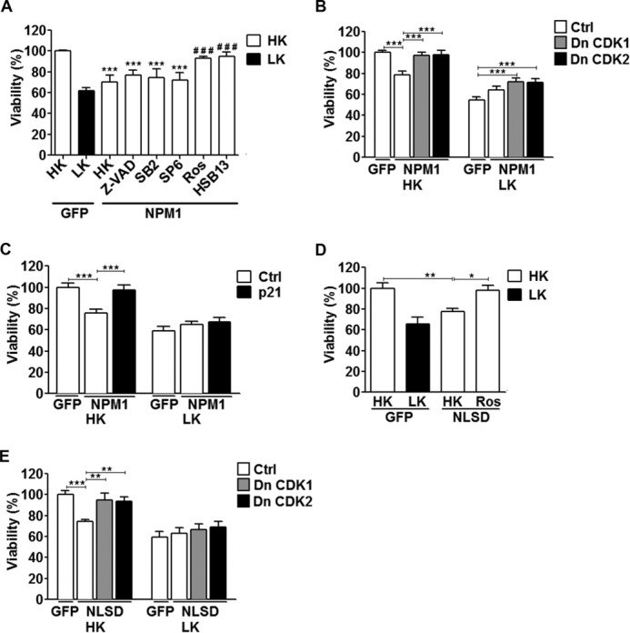
Toxicity by increased NPM1 expression is cell cycle-dependent. A, CGNs transfected with either EGFP or NPM1 were then treated with either HK medium alone or HK medium supplemented with the following inhibitors: Z-VAD (50 μm), SB216763 (5 μm), SP600125 (10 μm), roscovitine (50 μm), or HSB13 (25 μm). Viability was quantified by immunocytochemistry with a GFP or FLAG antibody. ***, p < 0.001 as compared with GFP in HK; ###, p < 0.001 as compared with NPM1 treated with HK (n = 3). B and C, CGNs were transfected with either EGFP and a control vector (Ctrl, pK3HA) or NPM1 and pK3HA (Ctrl), DnCDK1, DnCDK2 (B), or p21 (C) in a 1:2 ratio followed by HK/LK treatment. Viability was quantified based on NPM1 fluorescence by immunocytochemistry with a GFP antibody. ***, p < 0.001 (n = 3). D, CGNs transfected with EGFP or NLSD. The cells were then treated with either HK medium alone or HK medium supplemented with roscovitine (50 μm). Viability was quantified as described for A. *, p < 0.05; **, p < 0.01 (n = 3). E, CGNs transfected with EGFP and pK3HA (Ctrl) or NLSD and pK3HA (Ctrl), DnCDK1, or DnCDK2 in a 1:2 ratio followed by HK/LK treatment. Viability was quantified as described for B and C. **, p < 0.01; ***, p < 0.001 (n = 3). Ctrl, control.
