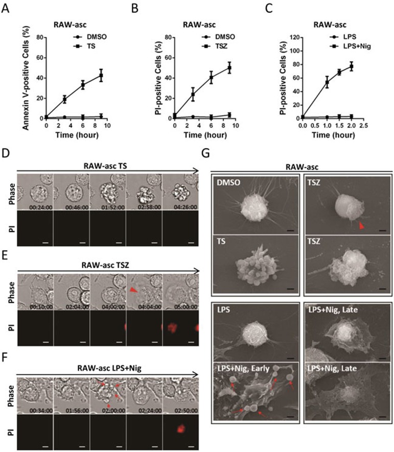
Figure 1.
Pyroptotic and necroptotic cells have distinct morphological features. (A-C) Viabilities of RAW-asc cells treated with DMSO or TS (TNF and smac mimetic, A), or TSZ (TNF, smac mimetic and the caspase inhibitor z-VAD, B), or primed with LPS for 4 h followed by nigericin (Nig) treatment or no treatment (C) for different periods of times as indicated. Apoptotic cells were identified by annexin V staining. Necroptosis and pyroptosis were determined by propidium iodide (PI) staining. Results shown are mean ± SD from three independent experiments. The final concentrations of 10 ng/ml TNF, 100 nM smac mimetic, 20 μM z-VAD, 1 μg/ml LPS and 10 μM Nig were used here and in subsequent experiments unless otherwise stated. (D-F) Representative time-lapse images of RAW-asc cells treated as in A-C. Cell morphology was visualized by wide-field light microscopy (upper panel) and cell membrane integrity was monitored by PI uptake (lower panel). (G) Representative scanning electronic microscopy (SEM) images of RAW-asc cells treated as in A-C. Arrowhead indicates explosion of necroptotic cells and arrow points to bubbling of pyroptotic cells. Scale bar, 5 μm (D-F) and 2 μm (G).

