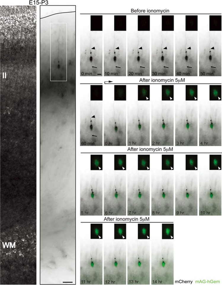
Figure 5.
Calcium influx induces mAG-hGem expression but not cell division in developing neurons from layer II at P3. Neuron with apical dendrite and axon (arrowhead and arrow respectively, in the time-lapse sequence) in layer II from P3 coronal brain slice (left panels, in utero electroporated at E15 with mCherry and mAG-hGem plasmids) express the S/G2/M marker mAG-hGem (white arrowheads, insets from time-lapse sequence) upon ionomycin treatment (5 μM). Scale bar: 50 μm (left panel) and 10 μm (time-lapse sequence).

