Figure 7.
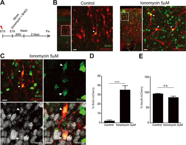
Calcium influx in developing neurons induces a limited proliferative potential. (A) Diagram depicting the steps followed during the experiments. (B) Control neurons in the CP are negative for BrdU and display a radial organization. Slices treated with ionomycin (5 μM) and washout for 2-3 days, contain mCherry-transfected cells in the CP positive for BrdU without a radial organization (white arrowheads, inset from the left panel). (C) Slices treated with ionomycin (5 μM) and washout for 2-3 days contain mCherry-transfected cells in the CP, positive for BrdU and NeuN (white arrowheads, inset from the left panel) that lost radial dendritic organization. (D) Quantification of mCherry-transfected cells in the CP positive for BrdU (***P = 0.0003 by t-test; values are mean ± SEM). (E) Quantification of mCherry-transfected cells in the CP positive for NeuN (P = 0.0814 by t-test; values are mean ± SEM). Scale bar: 10 μm.

