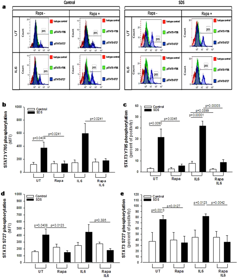
Figure 3. Flow cytometric analysis of STAT3 Y705 and S727 phosphorylation in LCLs.
Representative experiment indicating STAT3 Y705 (green histogram) and S727 (blue histogram) phosphorylation level in: (a) healthy donor derived LCLs (Control) versus SDS LCLs (SDS). Red histogram indicates isotype control. Control LCLs and SDS LCLs were pre-incubated with 350 nM rapamycin (Rapa) for 1 hour before stimulation in the presence or in the absence (UT) of IL-6 (10 ng/ml) for further 15 min. (b) Median Fluorescence Intensity (MFI) and Percent of positive cells (c) for STAT3 Y705 signal. (d) Median Fluorescence Intensity (MFI) and Percent of positive cells (e) for STAT3 S727 signal. Data are mean ± SEM of five independent experiments performed in LCLs derived from five different SDS patients. Student’s t-test has been calculated.

