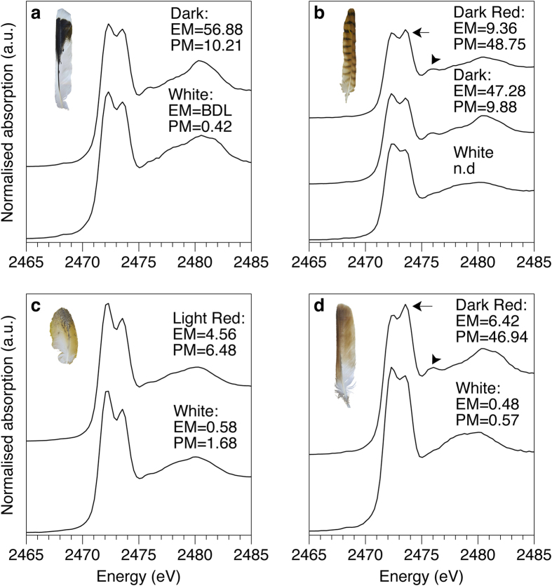
Figure 3.
Sulfur XANES of (a) Harris hawk, (b) kestrel, (c) barn owl, and (d) red-tailed hawk. All feather spectra are dominated by the characteristic disulfide double peak originating from keratin (compared to oxidised glutathione standard, Supplementary Fig. 7). The shift in peak intensities in pheomelanised tissues are indicated by arrows (2473.5 eV) and arrowheads (2476 eV). The identity of this shifted peak is best explained by the presence of a benzothiazole type (5-membered benzo-sulfur) units within the pheomelanin structure (Supplementary Fig. 7). EM = eumelanin μg/mg, PM = pheomelanin μg/mg, BDL = below detection limits, n.d. = no data.

