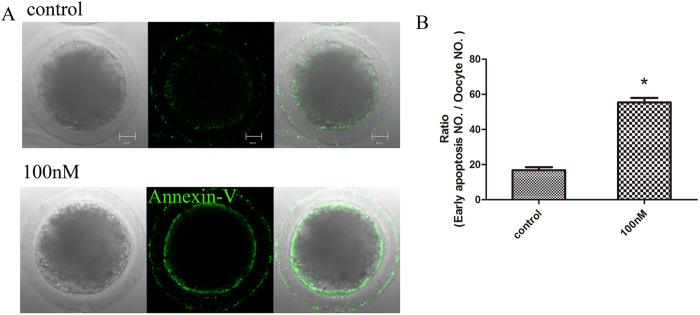
Figure 4. Early apoptosis was occurred among MI stage after HT-2 toxin exposure in porcine oocytes.
(A) Oocytes were stained with a FITC-conjugated Annexin-V. In control oocytes there were no fluorescent signals on the zona pellucida, whereas in HT-2 treated porcine oocytes, early apoptosis occurred and fluorescent signals were observed on the membrane. Annexin-V, green. (B) Percentages of oocytes exhibiting early apoptosis. The rate of oocytes with early apoptosis after HT-2 treatment was significantly increased compared with the control group. *p < 0.05. Bar = 20 μm.

