Figure 5.
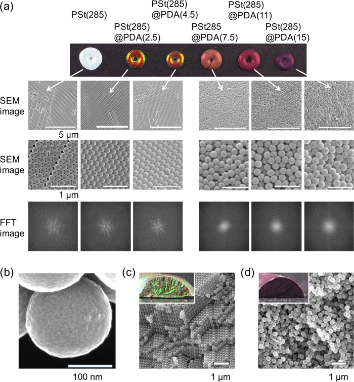
(a) SEM images and FFT spectra from SEM images of structural color pellets prepared by PSt(285)@PDA(Y) core-shell particles. FFT images were obtained from lower SEM images. (b) High resolution FE-SEM image of PSt(285)@PDA(15) particles. (c) Cross section view of PSt(285)@PDA(2.5) pellets. Inset shows a photograph of the samples. (d) Cross-section view of PSt(285)@PDA(15) pellets. Inset shows a photograph of the samples.

