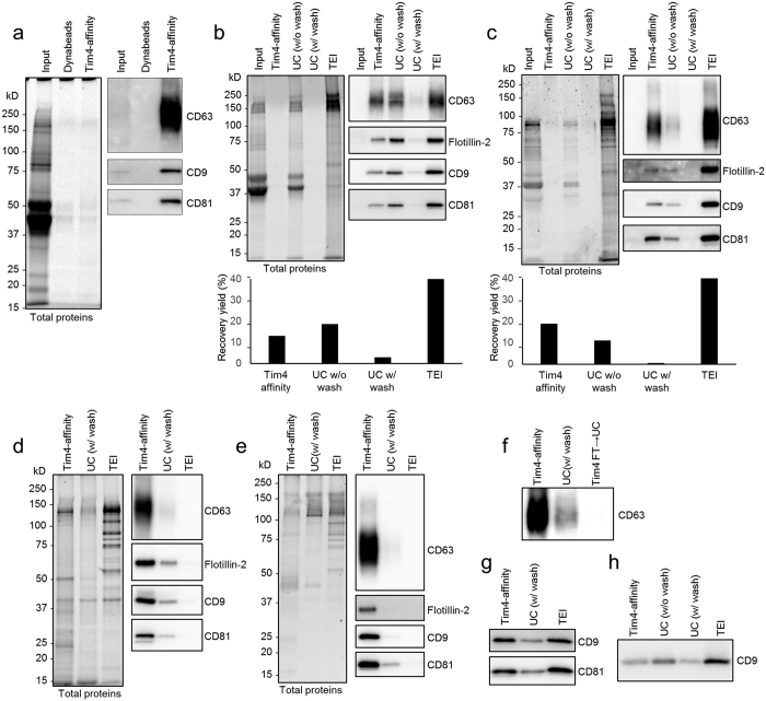
Figure 2. Tim4-affinity purification of sEVs.
(a) Thioglycollate-elicited mouse peritoneal macrophages (pMac) (3 × 10⁵ cells/ml) were cultured in 10 ml of DMEM containing 5% EV-depleted FBS for 48 h. 10K sup of pMac (4 ml each) were incubated with Dynabeads or mouse Tim4-Fc-conjugated Dynabeads overnight. Bound sEVs were eluted, subjected to SDS-PAGE and analyzed by western blot (right panel) with anti-CD63, anti-CD9, or anti-CD81 antibody. Total proteins in the sEV fractions were visualized by Oriole fluorescent gel stain (left panel). The amount loaded in the input lane was equivalent to 1/1000 volume of the total supernatant. (b,c) sEVs were purified from 10K sup (4 ml of medium containing 5% EV-depleted FBS) of K562 cells (1 × 10⁶ cells/ml) (b) or pMac (4 × 10⁵ cells/ml) (c) by each method. The purified sEV fractions were adjusted to the same volume (60 μl), subjected to SDS-PAGE, and analyzed by Oriole stain (left panels) or western blot (right panels). TEI: Total Exosome Isolation. The number of sEV particles in the purified fractions were also analyzed by NTA assay using NanoSight LM10 (Malvern), and the calculated purification recovery yield relative to total sEV particles was plotted in the lower graphs. (d,e) Protein concentration of purified sEV fractions from K562 cells (d) or pMac (e) was determined by BCA protein assay, and adjusted to the same in each method. Equal amount of proteins (100 ng) were subjected to SDS-PAGE, and analyzed by Oriole stain (left panels) or western blot (right panels). (f) The flow through of 10K sup of K562 cells after Tim4-affinity purification was further ultracentrifuged, and the same proportion of the protein amount to the total purified protein amount as Tim4-affinity purification was loaded to detect residual exosomes by western blot with anti-CD63 antibody (Tim4 FT → UC). (g,h) sEVs were isolated from 50 μl of mouse serum (g) or 500 μl of human urine (h) by each method, adjusted to the same volume and analyzed by western blot.

