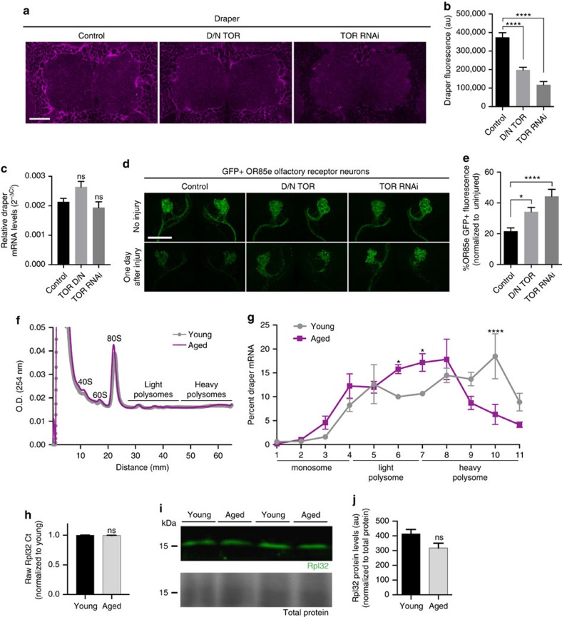
Figure 6. Inhibition of TOR in adult glia inhibits Draper and glial clearance of axon debris in young animals.
(a) Single confocal slices of antennal lobe regions. Brains immunostained with Draper. (b) Quantification of cortical Draper immunostainings shown in a; mean±s.e.m. plotted; ****P<0.0001, one-way analysis of variance (ANOVA) with Sidak post-hoc test; N≥16. (c) Quantitative PCR (qPCR) analysis of draper-I transcript levels in adult heads; mean±s.e.m. plotted, NS, not significant, one-way ANOVA with Sidak post-hoc test; N=3 biological replicates. (d) Representative Z-stack projections of OR85e GFP+ axons. (e) Quantification of GFP+ axonal debris shown in d; mean±s.e.m. plotted; *P<0.05 and ****P<0.0001, one-way ANOVA with Sidak post-hoc test. N=22. (f) Polysome profiles for young and aged whole head lysates resolved on a 10–60% sucrose gradient. Absorbance continuously monitored at 254 nm during fractionation shown. (g) draper-I mRNA in each fraction was quantified by qPCR and normalized to the housekeeping gene Rpl32 and luciferase (control for RNA recovery). mean±s.e.m. plotted; *P<0.05 and ****P<0.0001, two-way ANOVA with uncorrected Fisher's LSD post-hoc test. N=3 groups of 30 w118 fly heads/age. (h) Normalized Rpl32 Ct values pooled from four experiments on young and aged w118 brain lysates; mean±s.e.m. plotted, NS, not significant, unpaired t-test. N=17 biological replicates/age. (i) Representative western blotting for Rpl32 (green) and MemCode total protein stain (bottom panel) performed on head lysates from young and aged flies. (j) Quantification of Rpl32 western blottings; unpaired t-test. N=4 biological replicates/age. Genotypes: a–e, Control=w1118;OR85e-mCD8::GFP, tubulin-Gal80ts/+; repo-Gal4/+. D/N TOR=w1118;OR85e-mCD8::GFP, tubulin-Gal80ts/+; repo-Gal4/UAS-D/N TOR. TOR RNAi=w1118;OR85e-mCD8::GFP, tubulin-Gal80ts/+; repo-Gal4/UAS-TORRNAi. f–j, w1118. Scale bars=30 um.

