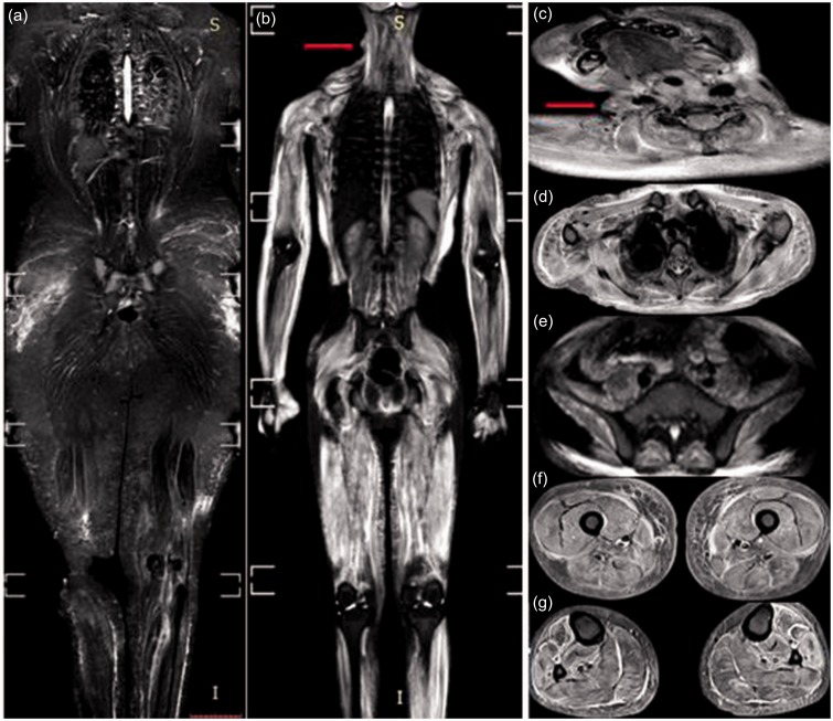
Fig. 1.
WB-MRI, STIR sequence for two cases of dermatomyositis. (a) Case 1: mild bilateral symmetrical edema of the gluteal, thigh, and calf muscles with interstitial edema of the subcutaneous fat of the gluteal region and thighs. Case 2: (b) marked bilateral symmetrical edema of all muscle groups: neck (c), the shoulder girdle and thoracic wall (d), the pelvic girdle (e), the thigh (f), and calf (g) muscles. Marked interstitial edema is clearly evident in the upper arms and the lateral aspect of the chest wall. Note: a skin nodule is seen on the right lateral aspect of the neck (arrow in b, c).

