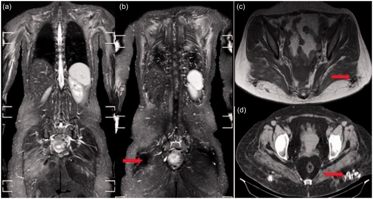
Fig. 4.
Overlap myositis with scleroderma in a 39-year-old woman. Bilateral symmetrical edema of the shoulder girdle and gluteal region (a), subscapularis, latissmus dorsi, and erector spinae muscles (b). Bilateral signal void foci are seen in the posterior gluteal subcutaneous fat on both coronal STIR (b) and axial T1 (c) images. They appear as hyperdense foci of calcification on the CT scan (d).

