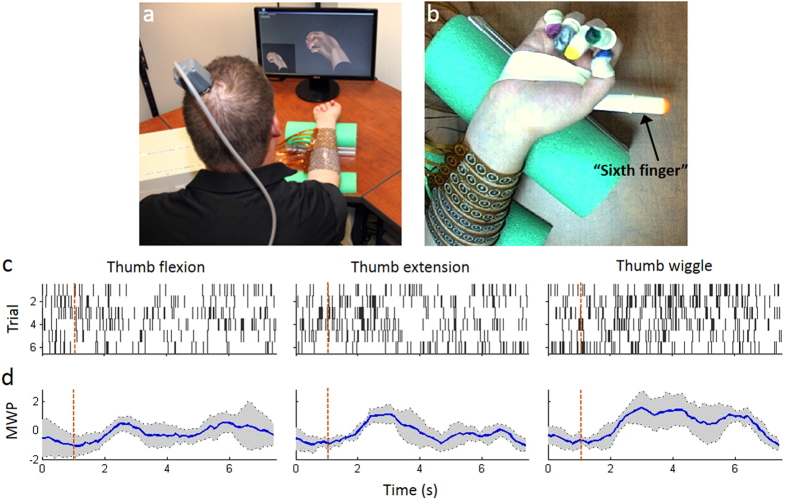
Figure 1.
(a) Experimental neural bypass system in use with the participant (seated in a wheelchair) in front of a table with a computer monitor. The neuromuscular electrical stimulation sleeve is wrapped around the participant’s forearm; (b) Close up view of the participant’s hand showing the colored cots on the fingers. An additional cot was placed on a plastic cylinder extending out past the participant’s thumb (referred to as the “sixth finger”). A stereo camera was positioned above the participant’s hand to track movement in 3 dimensions. The color of the cot on the thumb was used to identify thumb movement and locate it in three-dimensional space with the “sixth finger” serving as the reference point; (c) Examples of rasters histograms from one discriminated unit (channel 7 unit 1) with response to attempted thumb movements. The participant was presented with cues to attempt thumb flexion, thumb extension, and rhythmic thumb wiggle. Each cue was presented for a random duration of 2.5–4.5 s followed by a random 2.5–4.5 s rest period. We presented 6 trials of each in random order. Each dot in the raster represents a spike, and each row of spikes represents data from one trial. All trials were aligned on cue presentation (time 1 s, red dashed line). (d) Wavelet processed neural data from channel 7 corresponding to the same movements in panel c showing the mean wavelet power (MWP) averaged over the trials in dark blue with a confidence interval of ±1 S.D. around the mean shown in grey.

