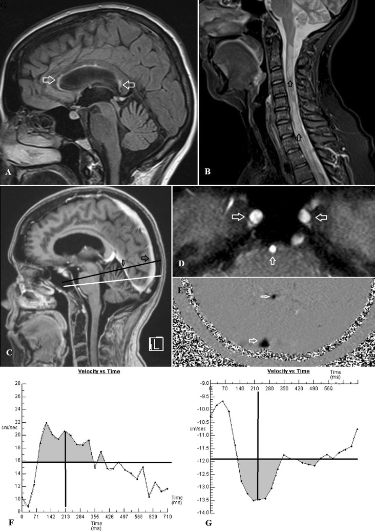
Fig. 1.

A A FLAIR sagittal image of a 48 year old female MS patient with arrows showing some plaques within the corpus callosum but no significant cerebral volume loss. B A T2 sagittal image of the cervical spine in the same patient with arrows showing extensive MS plaques. C A sagittal post contrast T1 image with the black line showing the position of the venous acquisition passing through the sagittal sinus (large arrow) and straight sinus (small arrow). The white line is the arterial acquisition passing through the skull base. D The localizer image from the arterial acquisition showing the carotid arteries just above foramen lacerum (horizontal arrows) and the mid basilar artery (vertical arrow). E The phase image from the venous acquisition showing the sagittal sinus (large arrow) and straight sinus (small arrow). F The arterial flow graph from the same patient. The horizontal line represents the mean flow velocity. Where the mean velocity transects the flow graph is the start and end of systole. The grey area above the line represents the arterial stroke volume which for the three arteries combined was 1184 µL. The vertical line represents the midpoint of the two transection points and is the midpoint of the primary arterial wave i.e. 213 mS. G The sagittal sinus flow graph from the same patient. The horizontal line is the mean flow velocity. The grey area is the sinus stroke volume which is 118 µL or 10 % of the arterial stroke volume. The vertical line is the midpoint of the two transection points and is the center of the primary pulse wave i.e. 223 mS giving an AVD of 10 mS in this patient which is less than 10 % of normal indicating very low compliance
