Fig. 3.

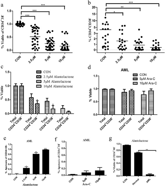
Alantolactone selectively induced apoptosis of LSCs in primary AML cells. a Alantolactone induced the apoptosis of CD34+CD38– cells with a dose-dependent manner in primary AML cells from AML specimens after treatment of alantolactone for 18 h. Each point represents an AML specimen. b Alantolactone reduced the percentage of CD34+CD38– cells with a dose-dependent manner in primary AML cells after the treatment of alantolactone for 18 h. Each point represents an AML specimen. c Alantolactone selectively induced apoptosis of CD34+CD38– cells comparing with total cells in primary AML cells after the treatment of alantolactone for 18 h by flow cytometry. d Ara-C showed negligible cytotoxicity to CD34+CD38– and total cells in primary AML cells at the concentrations of 5 and 10 μM. e Alantolactone induced apoptosis of CD34+CD38– cells in primary AML cells by flow cytometry at the concentrations of 2.5, 5, and 10 μM. f Ara-C showed almost no effect on inducing apoptosis of CD34+CD38– cells in primary AML cells at the concentrations of 5 and 10 μM. g Alantolactone treatment demonstrated significantly less effect on inducing apoptosis of CD34+CD38– cells in normal hematopoietic cells comparing to that in primary AML cells at a concentration of 10 μM. *P < 0.05, **P < 0.01, ***P < 0.0001
