Fig. 2.

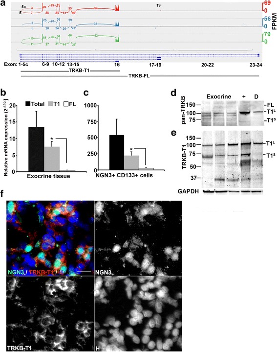
Quantitative analyses of TRKB isoform expression in cultured human exocrine tissue. a Isoform splicing of the neurotrophic tyrosine kinase receptor type 2 (TRKB) gene by NGN3+ cell populations isolated by coexpression of CD133 from 3 biological replicate exocrine cultures. TRKB loci and major isoforms shown below. Number of reads crossing exons are shown along with lines indicating joined exons. Height of peak indicates relative expression level in fragments per kilobase of transcript per million mapped reads (FPKM). Usage of alternate transcriptional start sites in exons 5 and 5c and alternate polyadenylation and stop codon in exon 19 are highlighted. b, c mRNA levels of total TRKB, TRKB-T1 and TRKB-FL isoforms. Relative Isoform expression calculated as mean ± SEM (n = 3 biological replicate cultures) of 2-ΔΔC t normalized to the level of cyclophillin A (PPIA) in (b) exocrine tissue and (c) NGN3+/CD133+ cells isolated from exocrine tissue. *, p < 0.05. TRKB levels in NGN3/CD133-depleted exocrine cells were too low to detect. d, e Western blot analyses of TRKB protein isoform expression. Protein lysates from 3 biological replicate exocrine cultures and NGN3+/CD133+ (+) and NGN3/CD133-depleted (D) cells from a single exocrine culture were probed with: d pan TRKB-specific TRKB antibody and (e) TRKB-T1-specific antibody. Apparent molecular mass for TRKB-FL, TRKB-T1 large size variant (T1L) and TRKB-T1 small size variant (T1S) shown at right. Molecular weight markers shown on left in kDa. Level of glyceraldehyde phosphate dehydrogenase (GAPDH) used as loading control. f Representative immunohistochemical staining of human cadaveric pancreas biopsy tissue for expression of NGN3 and TRKB-T1. Color overlay and individual monochrome images are shown with antibodies used. Nuclei counterstained with Hoechst 33342 (H). Scale bars are 20 microns
