
Fig. 9.
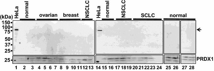
Immunoblotting of plasma exosomes for FEAT. Exosomes isolated from eight healthy volunteers and 18 cancer patients were analyzed by immunoblotting using rabbit anti-human FEATΔC (upper panel) and anti-peroxiredoxin 1 (PRDX1) (lower panel) antibodies. HeLa lysates from HeLa cells. The arrow denotes FEAT