FIGURE 3.
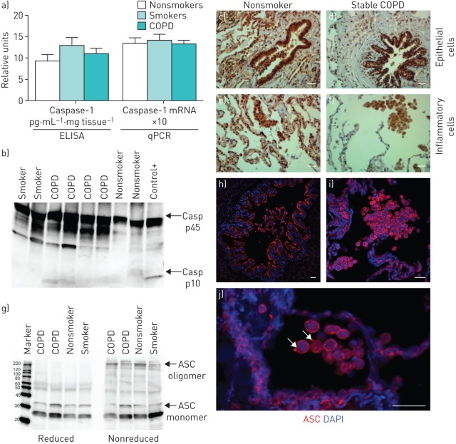
Inflammasome is not activated in stable chronic obstructive pulmonary disease (COPD). a) Caspase-1 protein levels measured by ELISA and relative mRNA expression measured by quantitative PCR (qPCR) in lung tissue of stable COPD. Data is presented as mean±sem of n=14 nonsmokers, n=15 smokers and n=38 COPD individuals. b) Representative Western blot analysis of caspase-1 (Casp); positive control (+) is a lysate of human peripheral blood mononuclear cells treated with the NLRP3 activator nigericin (10 μM). c–f) Caspase-1-positive cells (brown) detected by immunohistochemistry in c, d) bronchial epithelial cells and alveolar epithelial cells and e, f) macrophages in a representative tissue sample from a nonsmoker and a COPD patient. The primary antibody used in immunohistochemistry detects both the p45 inactive precursor and the small subunit of active caspase-1 (p10). Scale bars: 100 μm. g) Western blot for ASC in representative lung samples from two COPD patients, a nonsmoker (NS) and a smoker (S), denoting the monomeric (22 kDa) and oligomeric (220 kDa) ASC. ASC oligomeric complexes were not detected when samples were run under reduced conditions. h–j) ASC immunofluorescence staining (red) and nuclei (blue) in lung tissue of a representative COPD patient: h) alveolar epithelial cells and macrophages, i) bronchial epithelial cells, and j) macrophages. Arrows in j) denote macrophages with detectable ASC speck. DAPI: 4′,6-diamidino-2-phenylindole. Scale bars: 36 μm.

