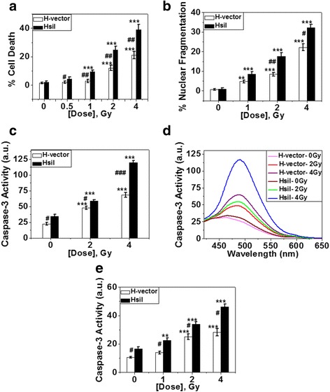
Fig. 5.

Study of different modes of cell death. a % of cell death as detected by trypan blue staining after 19 h of post-carbon ion exposure and plotted. Each bar diagram represents the mean ± SD of four independent experiments. b Detection of nuclear fragmentation. % nuclear fragmentation was calculated and plotted. Each bar diagram represents the mean ± SD of four independent experiments. Typical image of fragmented nucleus is given in Additional file 4a. c & d Assay of caspase-3 activity by fluorimetric study. Fluorimetric assay of caspase-3 activity of both H-vector and HsiI cells after 20 h of post-carbon ion exposure is shown here (c). Each bar diagram represents the mean ± SD of three independent experiments. The raw fluorescence spectra representing caspase-3 activity after carbon ion exposure is given here (d). e Caspase-3 activation by IF study. Fluorescence intensities of caspase-3 immunostained cells in 20 h post-carbon ion exposure were measured by ImageJ software and plotted. Each bar diagram represents the mean ± SD of three independent experiments. Typical photographs of immunostained cells are provided in Additional file 4b
