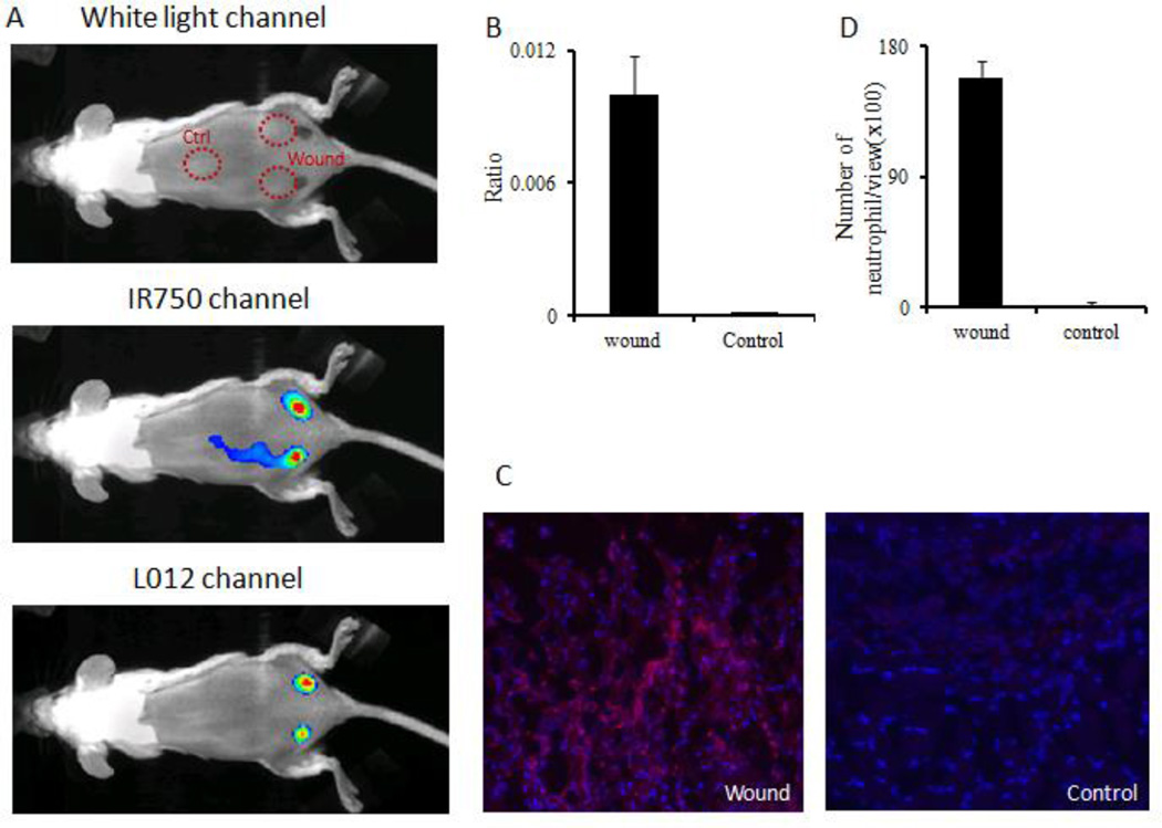
Figure 5.
ROS ratiometric probes were used to assess the extent of ROS activities on skin incision wounds. (A) White light/fluorescence/chemiluminescence images were taken from the animal 10 minutes after probe placement. (B) Quantification of chemiluminescence and fluorescence intensity ratios at the wound sites and control (uninjured) tissue. (C) Immunohistochemistry staining images taken on sections of wounded tissue and uninjured control tissue. (D) Quantification of neutrophil numbers in wound tissue and control tissue sections under microscope.

